Mole Mapping Technology
Detecting melanoma for early prevention.
While most moles do not become melanoma, they are the most common place for melanoma to form. Advanced imaging mole map technology can help detect new moles and compare changes in existing moles for early treatment and successful elimination of melanoma if detected.

Every eight minutes, someone in the U.S. will be diagnosed with melanoma.* The good news is that with early detection, melanoma has a cure rate of over 95%.
The Melanoma Research Foundation
Our smart skin imaging system.
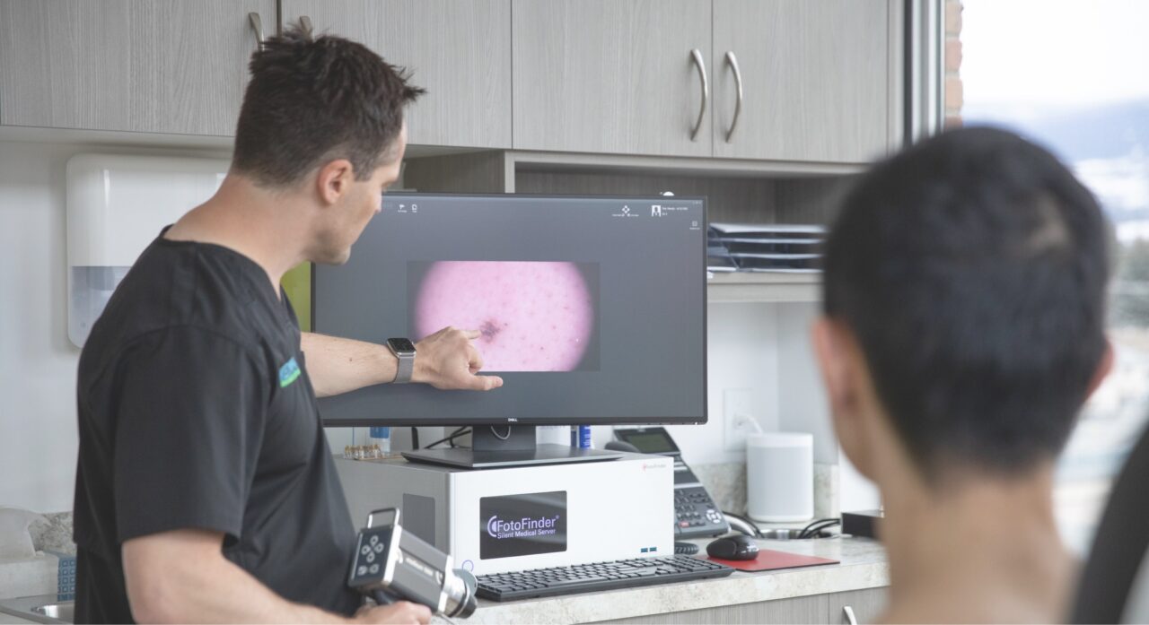
Dr. Ben Wiese uses the FotoFinder Automated Total Body Mapping (ATBM) to create an image database of all of your moles. As an AI (Artificial intelligence) based software, it allows for a proper evaluation of malignancy risk of melanoma. You can feel safer knowing that any changes or new moles will not be missed.
High-resolution photography
A clear and detailed picture gives you peace of mind in making the right decisions.
State-of-the-art software
Quicker and more advanced detection with the latest in diagnostic technology.
Health Canada approved
Your scans and personal information will be kept safe and secure.

How mole mapping works.
- FotoFinder is a Health Canada approved, computerized skin mapping system that creates accurate photos of your moles.
- The high-resolution camera is connected to a computer and transfers all photos directly to the doctor’s database.
- This whole-body mole check provides a mole risk score through the use of artificial intelligence for the assessment of lesions.
- Your doctor can compare your moles with photos from your initial visit and immediately identify any abnormalities.
Request an appointment.
Please don’t hesitate to get in touch with our office to experience direct access to our first-rate health services and patient-focused care.
Frequently asked questions
Digital mole mapping may be appropriate for people at a higher risk for melanoma. If you meet any of the criteria listed below, you are at a higher risk for melanoma:
- Multiple moles on your body (more than 50)
- A history of melanoma in your family
- A previous melanoma or skin cancer diagnosis
- A previous diagnosis of dysplastic moles
- Large (more than 2 millimetres in diameter) and/or oddly shaped moles
- Noticeably changing moles or new moles
- A history of severe, blistering sunburns during childhood or adolescence
- A history of tanning bed use
- Very light skin/pale complexion
In order to prepare for your mole mapping session:
- Wear comfortable clothing and shoes that are easy to take off.
- Preferably wear black underwear and remember you will need to wear similar undergarments for subsequent mole mapping sessions. Boxer shorts are not recommended.
- Wear as little jewelry as possible. (Remove watches, bracelets, necklaces, chains, earrings, and piercings, etc.)
- Please do not wear makeup, lipstick, eyeliner or nail polish.
- Do not use self-tanning products in the week prior.
- If you have long hair, please wear your hair up during the photography.
If you have moles that you would like mapped in areas covered by your undergarments, please discuss this with the medical assistant when you arrive for your appointment.
Full body photography in conjunction with a complete full body examination will typically take up to 60 minutes.
Digital mole mapping should be performed every 6 to 12 months as recommended by your doctor.
Please call our office at 236-420-3277 to schedule your privately paid digital Mole Mapping and complete full-body examination.
Melanoma is a skin cancer that commonly develops in melanocytes, the cells that produce melanin or pigment for the skin, hair and eyes. Melanocytes can also form moles, and while most moles do not become melanoma, they are also the most common place for melanoma to form. If a mole is changing it could be a sign of melanoma. Early detection of melanoma is very important as it has a cure rate of over 95% when found in its early stage.
One of our skin cancer trained nurses will take your photos with the FotoFinder Automated Total Body Mapping system. Your physician will review your photographs, analyze any changes and discuss them with you during your full-body skin exam.
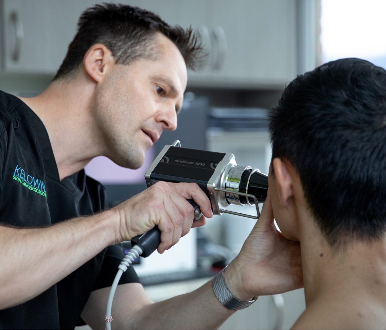
You will undress to your level of comfort and be positioned in front of the FotoFinder camera. Firstly full body overview photographs will be taken. At this point, Dr. Wiese will do a full body skin exam with a handheld device called a dermatoscope and will mark any moles that need magnification photography and will then take close-up photos of these moles with a handheld device.
You cannot have digital mole mapping performed when you have a rash, sunburn or prominent tan lines as this can interfere with the detection software.
Digital mole mapping with FotoFinder is only an aid for your doctor. You will still need to get full-body skin exams. Although the digital mole mapping is very helpful, the software may not detect all new or changing lesions. Full-body skin exams are very important, and they are not replaced by digital mole mapping.
Using the “ABCDEFG” rule can help you to recognize suspicious moles during self-evaluation. Moles which show one or more of the signs below should be treated with the utmost attention and observed by your physician!
- A for Asymmetry
- B for irregular, Blurred or jagged Borders
- C for Colour variation
- D for Diameter larger than ¼ inch
- E for Evolving, any change – in size, shape, colour, elevation, or another trait
- F for Firm Feeling
- G for Growing in size
Scientific studies on mole mapping.
Indications for Digital Monitoring of Patients with Multiple Nevi
Recommendations from the International Dermoscopy Society
Russo et al. (2022)
The Value of Total Body Photography for the Early Detection of Melanoma
A Systematic Review
Hornung et al. (2021)
The importance of total-body photography
Sequential digital dermatoscopy for monitoring patients at increased melanoma risk
Deinlein et al. (2020)
Usefulness of the ‘two-step method’
Digital follow-up for early-stage melanoma detection in high-risk French patients: a retrospective 4-year study
Gasparini et al. (2019)
Benefits of total body photography and digital dermatoscopy
Early diagnosis of melanoma in patients at high risk for melanoma
Salerni et al. (2012)
Total body skin examination
Skin cancer screening in patients with focused symptoms
Argenziano et al. (2012)
