MELANOMA
The most severe form of skin cancer.
Melanoma is connected to melanocytes, the cells that secrete melanin pigment and are responsible for skin colour. When the skin gets exposed to the sun, melanocytes produce more melanin.

What is melanoma?
This type of cancer can develop anywhere on the skin. While melanoma can often be found on the neck, head, and back in men, it usually occurs on the lower part of the legs and the back in women. It’s danger can be attributed to the fact that it can spread to other body parts.
The seven common types of melanoma cancer
Superficial Spreading Melanoma

Overview: Superficial spreading melanoma appears like a black or dark brown mark, which spreads from a new or an existing mole. It is generally in the skin areas that are exposed to UV rays, particularly in regions with a previous sunburn. As the most common type, it makes up for 70% of all melanomas.
Appearance: Typically, these lesions are over 6 mm and often 10+ mm in diameter. They will be irregular, asymmetric, raised, and multicoloured. They are often found on the torso of males and the legs of females, as well as the upper back of both sexes.
Dermoscopy Features: Chaotic in appearance, with more than one pattern and/or more than one colour arranged asymmetrically. They are often multicoloured. They can be blue or grey structures, lines radial, or pseudopods segmental, peripheral black dots or clouds, white lines, and polymorphous vessels.
Lentigo Maligna Melanoma

Overview: Lentigo maligna melanoma looks like a dark mark with an irregular border and uneven colour. It might initially appear like an irregular, large freckle, and is often found on the arms or face of older and middle-aged people.
Appearance: These lesions are often large, over 6 mm in size, have an irregular shape and a smooth surface. They have variable pigmentation, light brown, dark brown, pink, red, or white. Lesions will increase in size over several years. Ulceration, bleeding, itching, or stinging can occur. Thus, increasing numbers of colours and thickening part of the lesion.
Dermoscopy Features: Circles can appear brown or grey, multicoloured, light brown to black, and pigmented.
Nodular Melanoma

Overview: Nodular melanoma looks like a domed, rigid bump, and it is black or dark brown in colour. Nodular melanoma can spread rapidly from the epidermis to the dermis. Once it reaches the dermis, it can spread further to other parts of the body. It contributes to 10% of all melanomas.
Appearance: These lesions present as a nodule or papule and may be symmetrical in all directions. The nodules can have a variety of skin colours, tan, blue, or red. Their surface may be smooth, rough, crusty, or warty. The tissue can be very fragile and bleed on contact, or it can also be asymptomatic. It may appear on any site, but most commonly occur on exposed areas of the head and neck.
Dermoscopy Features: Multicoloured and chaotic in appearance, with more than one pattern or more than one colour arranged asymmetrically. Some dermoscopy clues to this type of lesion include grey and blue structures, polarizing specific white lines and polymorphic vessels.
Melanoma Desmoplastic

Overview: Desmoplastic melanoma is a rare type of melanoma. It occurs in sun-damaged parts of the skin, usually in older people. It appears like a slow-growing irregular or round lump. It is often scar-like and rigid with uneven colouration.
Appearance: These lesions can vary in size from medium to large, and appear as a raised indurated papule, nodule or plaque. The border is large and poorly demarcated, and lesions are skin coloured to pink. Symptoms can include ulceration, bleeding, itching, or stinging.
Dermoscopy Features: Structureless area (scar like) with grey dots. Appears brown, tan, white, and milky red. Some dermoscopy clues to this type of lesion include regression structures, with scar-like appearance. Polarized dermoscopy reveals white lines. Vascular structures, including serpentine vessels, dotted vessels and or milky red areas.
Melanoma Acral Lentiginous

Overview: Acral lentiginous melanoma usually appears like a bruise or dark mark that doesn’t heal. It can occur anywhere, including the soles of the feet and palms of the hands. Under the nails, this type of melanoma might appear like a dark stripe. It is quite uncommon.
Appearance: Size varies, depending on when diagnosis is made. It can vary from a few millimetres to several centimetres. Appears as a patch or macule that is initially difficult to recognize, but later becomes irregular in shape and colour. Mixture of brown, blue-grey and red colours. Typically appear on palms, soles, fingers, and toes.
Dermoscopy Features: Parallel ridge pattern with chaotic, multicoloured appearance. Some dermoscopy clues to this type of lesion include grey and blue structures, lines parallel, ridges or chaotic, black dots, or clods peripheral.
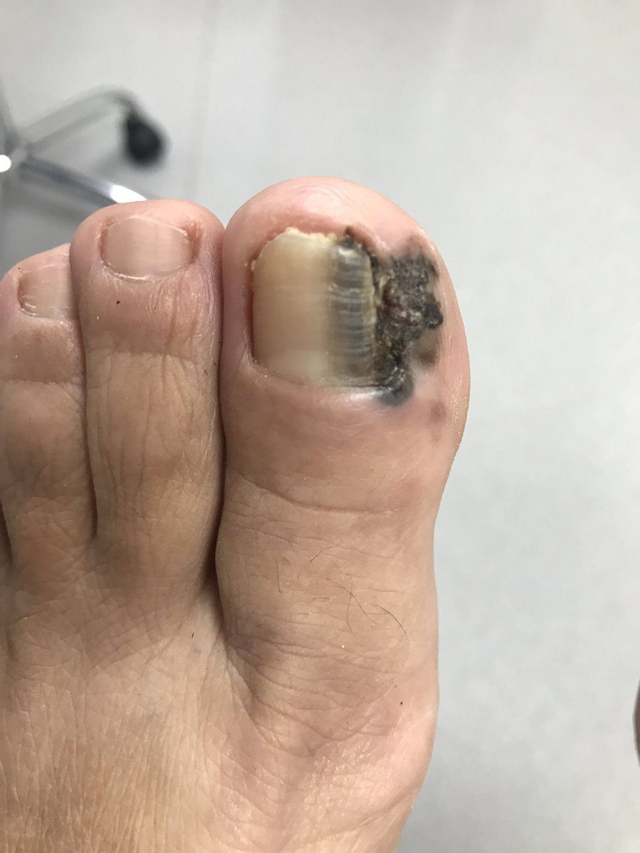
Subungual Melanoma

Overview: Subungual melanoma is also a rare form of melanoma, it develops under the nails of the fingers or toes. It usually occurs in individuals with darker skin tone. Often mistaken as a bruise, it is black or brown in appearance.
Appearance: Typical size is over 3mm new or changing band. Parallel pattern of lines, varying colours, brown, grey, yellow, or black. They either have no surface features, or have nail plate splitting or roughness, unless advanced with nodular lesion palpable. Can appear on any finger or toenail. Typically, asymptomatic, but can progress, ulcerate or bleed, or severe pain with bone involvement.
Dermoscopy Features: Chaotic, parallel lines with grey and black.
Uveal melanoma

Overview: uveal melanoma is a type of eye cancer that involves the choroid, ciliary body, and iris. The tumour develops from the melanocytes lying within the uveal region.
Diagnosis of melanoma.
Your doctor will cut out a lesion that is suspicious for melanoma. The tissue is then sent to a laboratory to be analyzed by a pathologist, who examines the sample under a microscope for melanoma diagnosis. If melanoma is confirmed, an additional surgical procedure may be required, depending on the depth of the lesion and several other factors. Thankfully, in most cases of melanoma, no further treatment is needed.
Diagnostic tests for melanoma include: blood tests, image testing and surgical tests.
Staging and Treatment of Melanoma Skin Cancer
On the basis of the reports of imaging and surgical tests, the doctor tries to find if the cancer has spread and to what extent. Read our helpful article for information on the stages of melanoma and corresponding treatments.
Who is most at risk?
Anyone can develop melanoma. However some of the more common risk factors include:
- Fair Skin Tone
- Excessive Sun Exposure
- Sunburn History
- Use of Tanning Beds
- History of Precancerous Skin Lesions
- History of Skin Cancer
- Weakened Immunity
Preventing melanoma.
Full body protection
Protect any part of the body that is not covered with UV protective clothing. Use a broad spectrum UVA/UVB sunscreen with a minimum SPF of 30-45.
Develop sun-safe habits
Avoid harsh UV rays and seek shade between the sun’s peak hours from 10 am to 4 pm. Or simply, stay out of the sun.
Avoid tanning
Prolonged UV exposure from tanning beds can lead to as much damage as sun exposure can. Tanning bed use over time can also lead to actinic keratosis. There is no such thing as a healthy tan, or safely “pre-tanning” before going on a holiday to a sunny destination.
Check your skin regularly
Keep an eye out for any changes in spots, freckles, blemishes, or abnormal skin growths. If they hurt, bleed or grow over time, see your doctor.
Get to know your treatment options.
Basal cell carcinoma treatments are available through our clinic, including:Melanoma treatments available through our clinic. Due to the severity of melanoma, treatments can vary exponentially. Your doctor will advise on your best course of action.
Talk to a physician.
With all types of skin cancer, it’s important to seek the advice of a qualified skin health physician. At Kelowna Skin Cancer Clinic, we have tools and expertise to diagnose and guide you through your treatment options.
