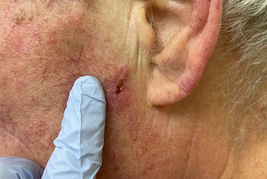
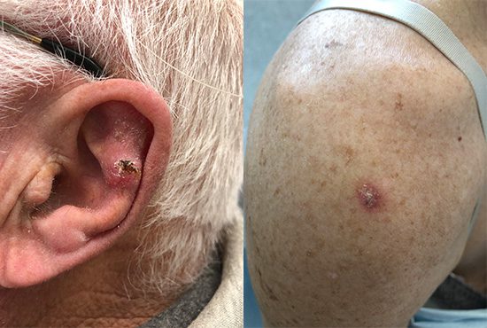
SQUAMOUS CELL CARCINOMA
Common and easily to identify.
Squamous cell carcinoma (SCC) of the skin is the second most common type of skin cancer. This skin cancer develops in the squamous cells that form the outer and middle layers of the skin. If detected early, most SCCs are treatable.
Though it’s not usually deadly, it can be aggressive. If left untreated, it may get bigger and even spread to other body parts, resulting in serious health complications.

What is squamous cell carcinoma?
Squamous cells are one of three types of cells in the epidermis. They are flat and found near the skin’s surface. These squamous cells shed constantly with the formation of new ones. SCC happens when damage to the DNA occurs, usually due to exposure to harmful ultraviolet rays or any other agents that may trigger DNA damage.
How to spot squamous cell carcinoma.
Squamous cell carcinomas of the skin look like open sores. They may itch, crust, or bleed. These lesions mostly develop on the sun-exposed areas of the skin. It may also occur on body parts like the genitals.
Signs & Symptoms
- Red, firm nodule
- Flat sore with scaly crust
- Any new sore or raised region over an old ulcer or scar
- Any scaly, rough patch on the lips that could develop into an open sore
- A reddish rough sore or patch inside the mouth
- Red, raised patch or wart-like structure over or inside the anus or on the genitals

Features of Each Subtype
SCC in Situ (Bowen’s Disease)
Appearance: These lesions often have an irregular border, appear red, pink, or reddish-brown and are scaly and crusty. They are typically asymptomatic but can itch and bleed.
Dermoscopy Features: The most common pattern is structureless and brown. Less commonly it will be an asymmetric combination of brown and/or grey dots, hypopigmented structureless area. Colours can be brown, grey, and occasionally structureless. Some dermoscopy clues to this type of lesion include dots arranged in line, coiled vessels.
SCC Well Differentiated
Appearance: The size can vary from a few millimetres to a few centimetres. It has a domed shape and is rough and raised. It appears pink and is distributed relatively evenly around the periphery of the lesion. It will typically appear on sun-exposed areas such as head, neck, upper extremities, but can occur anywhere on the body, including mouth, anus, and genitals. They can be tender and painful, as well as cause paresthesia and dysesthesia.
Dermoscopy Features: The pattern includes all types of vessels as lines, including coiled ones, as well as the occasional branched vessels. Colour appears as a whitish structureless area. Some dermoscopy clues to this type of lesion include surface keratin, white structureless areas and white circles.
SCC Poorly Differentiated
Appearance: The size can vary from a few millimetres to a few centimetres. They appear as papules or nodules. Appearance can vary from ill-defined patches to well-circumscribed hyperkeratotic plaques that are rough and raised. They have pink areas that are more frequently central and in an irregular spatial arrangement. They typically occur in sun-exposed areas. They will be tender and painful.
Dermoscopy Features: They are perifollicular white circles. All types of vessels as lines, including coiled ones, occasionally branched vessels. The vessels cover more than 50% of the lesion. They are pink in colour. Some dermoscopy clues to this type of lesion include absence of surface keratin, white structureless areas and white circles.
Who is most at risk?
Anyone can develop squamous cell carcinoma. However some of the more common risk factors include:
- ExposuFair Skin Tone
- Excessive Sun Exposure
- Sunburn History
- Use of Tanning Beds
- History of Precancerous Skin Lesions
- History of Skin Cancer
- Weakened Immunity
Preventing squamous cell carcinoma.
Full body protection
Protect any part of the body that is not covered with UV protective clothing. Use a broad spectrum UVA/UVB sunscreen with a minimum SPF of 30-45.
Develop sun-safe habits
Avoid harsh UV rays and seek shade between the sun’s peak hours from 10 am to 4 pm. Or simply, stay out of the sun.
Avoid tanning
Prolonged UV exposure from tanning beds can lead to as much damage as sun exposure can. Tanning bed use over time can also lead to actinic keratosis. There is no such thing as a healthy tan, or safely “pre-tanning” before going on a holiday to a sunny destination.
Check your skin regularly
Keep an eye out for any changes in spots, freckles, blemishes, or abnormal skin growths. If they hurt, bleed or grow over time, see your doctor.
Get to know your treatment options.
Basal cell carcinoma treatments are available through our clinSquamous cell carcinomas treatments available through our clinic. The most common treatment options would be the following:
Curettage & electrodesiccation, cryosurgery, and photodynamic therapy.
If the cancer has spread, your doctor may recommend more serious treatments, such as surgery or chemotherapy.
Talk to a physician.
With all types of skin cancer, it’s important to seek the advice of a qualified skin health physician. At Kelowna Skin Cancer Clinic, we have tools and expertise to diagnose and guide you through your treatment options.
