
DERMOSCOPY
Putting your skin under the lens
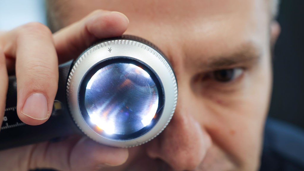
Skin cancer dermoscopy uses a handheld tool called a dermatoscope to examine the skin more closely. Its powerful magnifying lens lets clinicians see structures beneath the surface that aren’t visible to the naked eye. This enhanced view helps identify unusual changes earlier and supports timely diagnosis.
There are two types of dermoscopy
Non-polarized dermoscopy
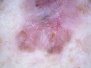
This method uses direct light to illuminate the skin. It’s helpful for viewing surface features and capturing clear images of the affected area.
Polarized dermoscopy
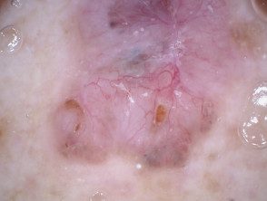
This method uses cross-polarized light to reduce glare and see deeper structures without altering how the lesion appears. It’s especially useful when a clinician needs a closer look at features beneath the surface.
Precision evaluation for your skin health
Concerned about changing spots or unusual lesions?