


BENIGN SKIN GROWTHS
Seborrheic keratosis
Common, benign skin growths
Seborrheic keratoses are common, non-cancerous skin growths that become more frequent with age. They often have a waxy, warty, or “stuck-on” appearance. Once the diagnosis is confirmed, treatment is usually unnecessary, although removal may be considered for irritation or cosmetic reasons.
Some skin cancers can look similar, so a new, changing, bleeding, or uncertain growth should be assessed rather than diagnosed from a photograph.
At a glance
Benign, not precancerous
A confirmed SebK is not a skin cancer.
Common with age
New growths may develop gradually over time.
Usually no treatment needed
Removal can be considered for irritation or appearance.
Assess uncertain changes
Some skin cancers can resemble SebK.
Appearance
Seborrheic keratoses have a classic “stuck-on” or “pasted-on” appearance — as if a small piece of wax or a scab were sitting on top of the skin.
Skin of colour: Small, dark, SK-like spots on the cheeks and temples are often a related condition called dermatosis papulosa nigra.
Causes
The exact cause is unknown. Seborrheic keratoses become more common with age, and some people have an inherited tendency to develop them. They are not contagious.
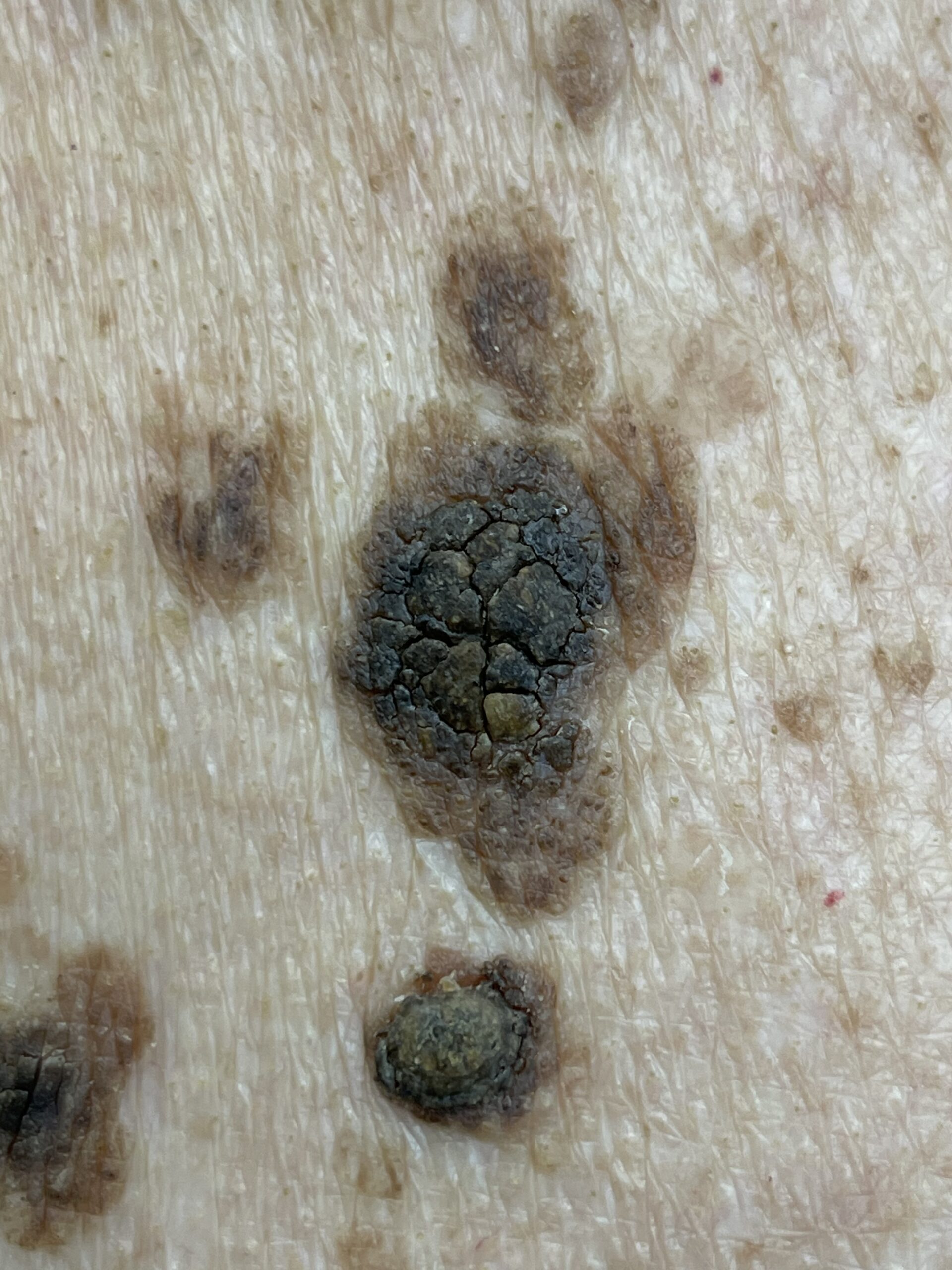
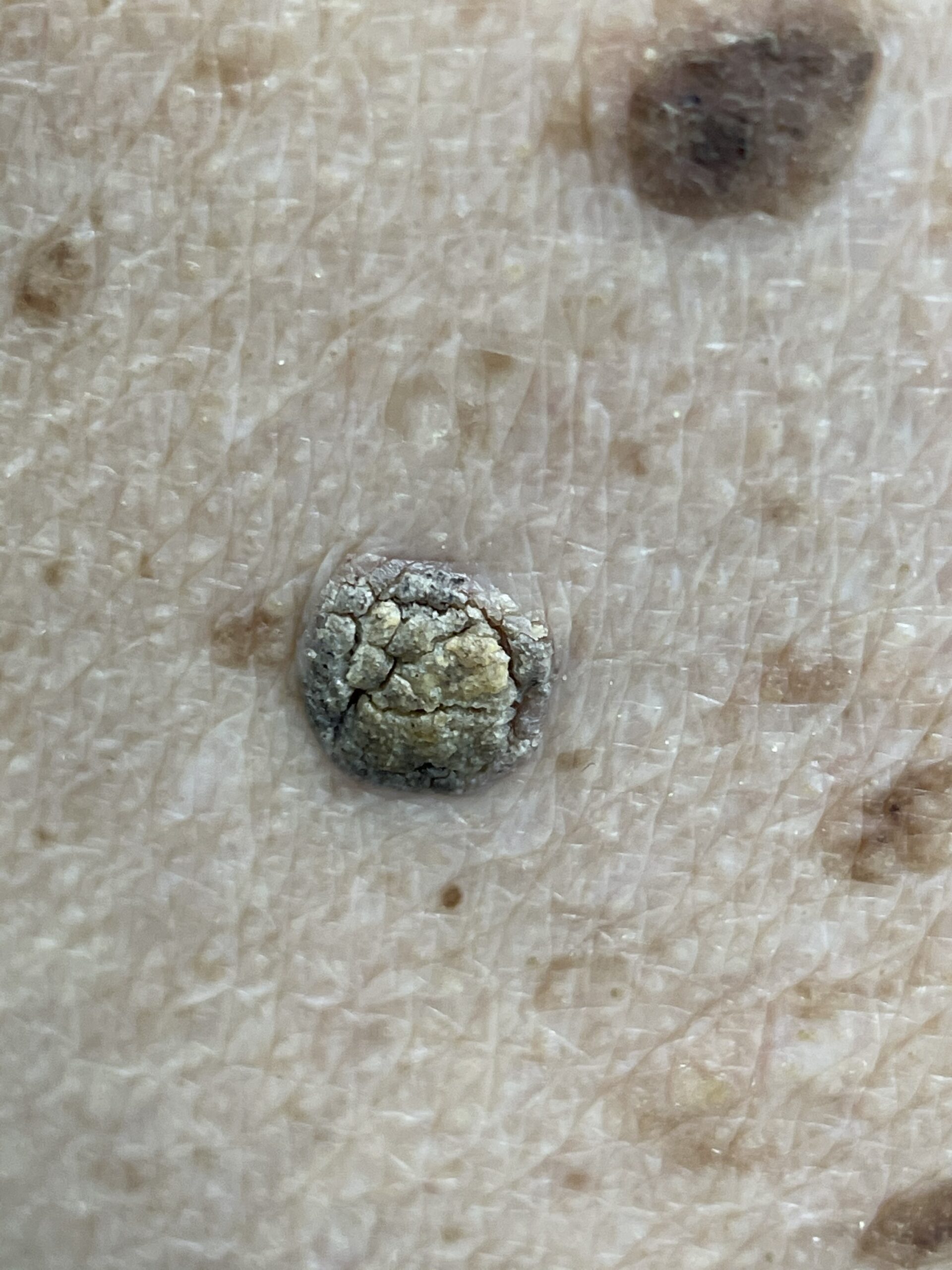
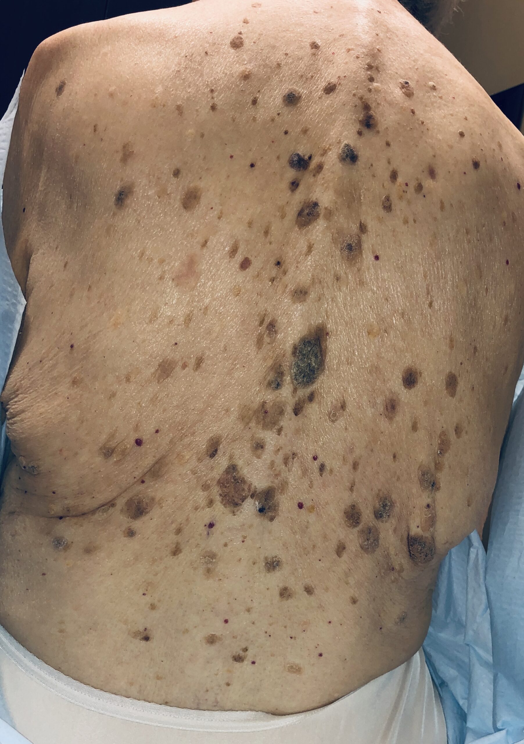
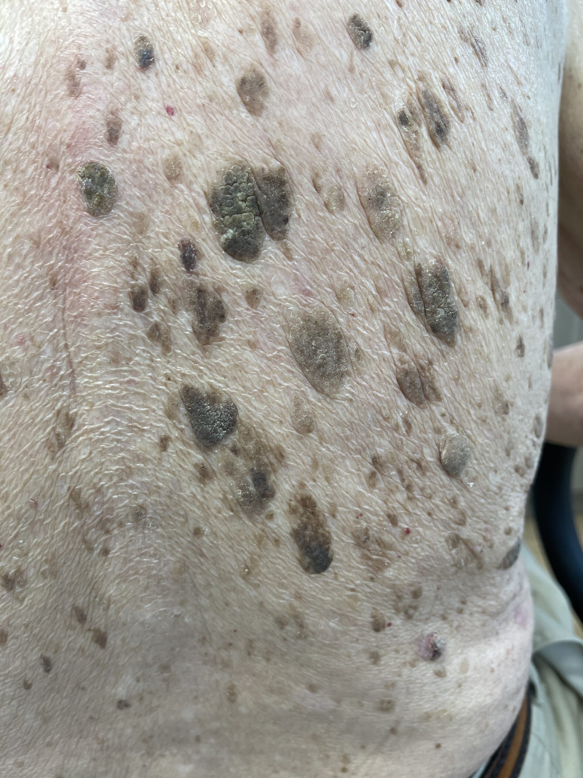
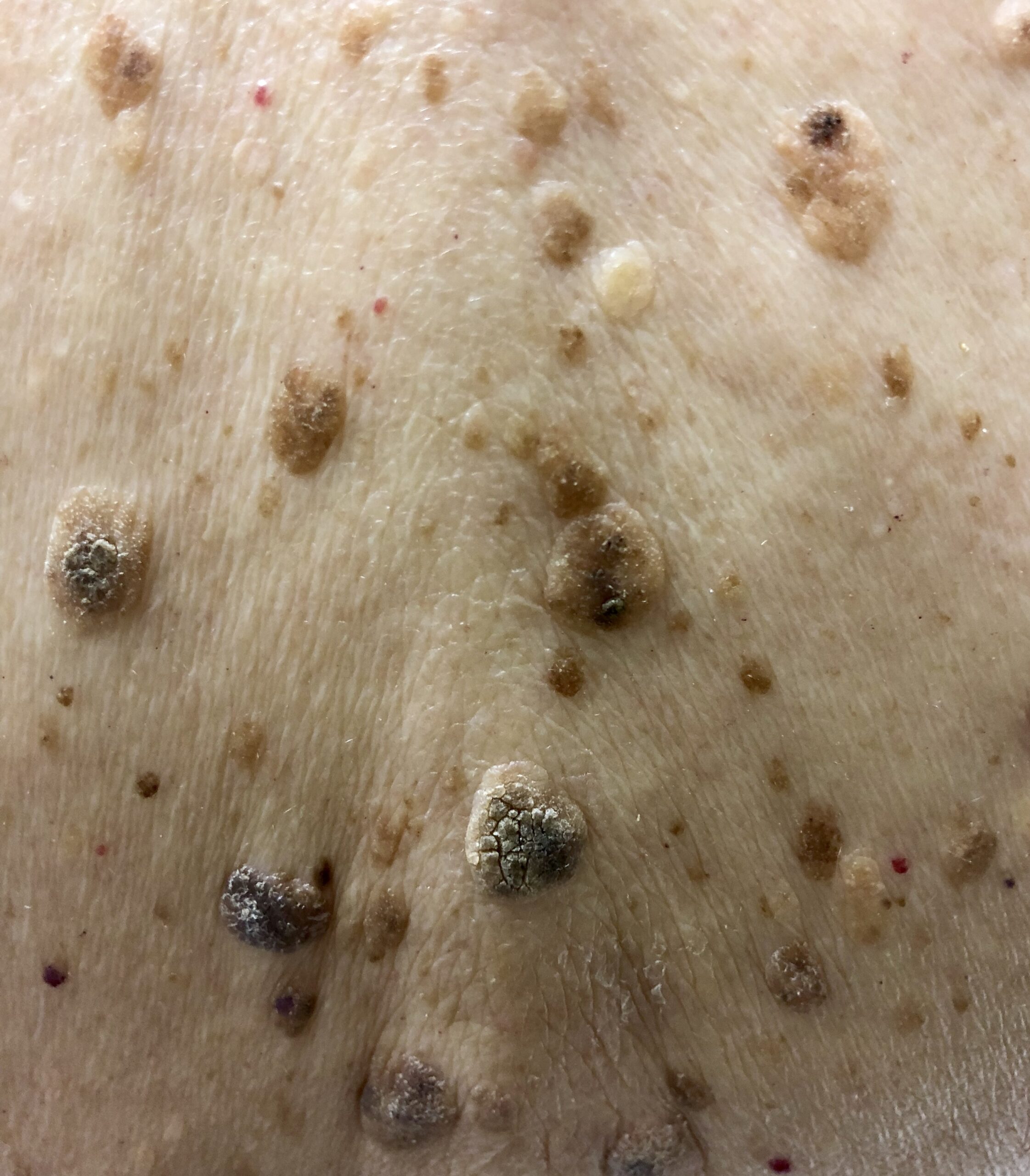
Clinical photographs of seborrheic keratoses
These clinical photographs show different colours, textures, thicknesses, and distributions. Matching a photograph is not a substitute for assessment.
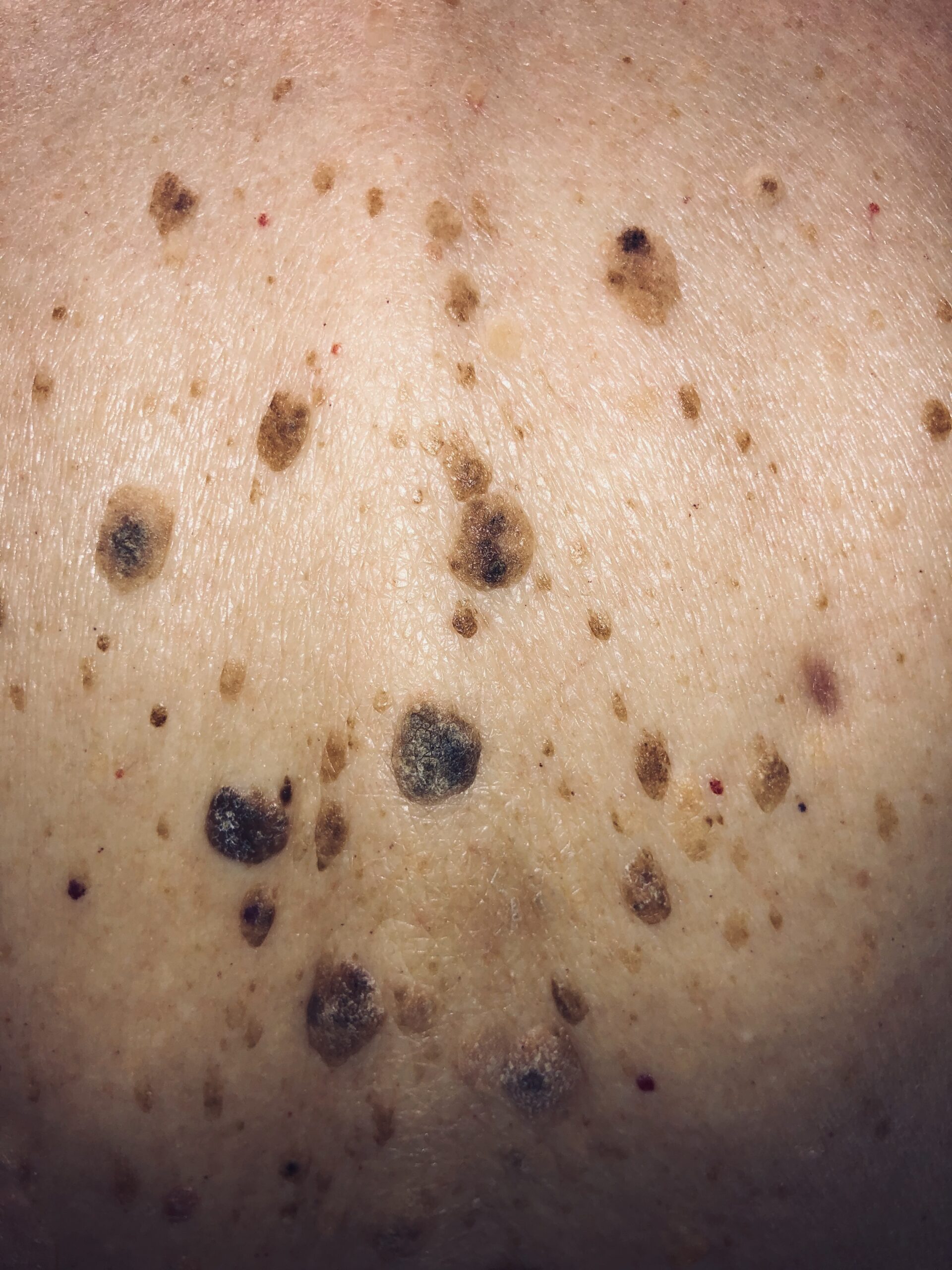
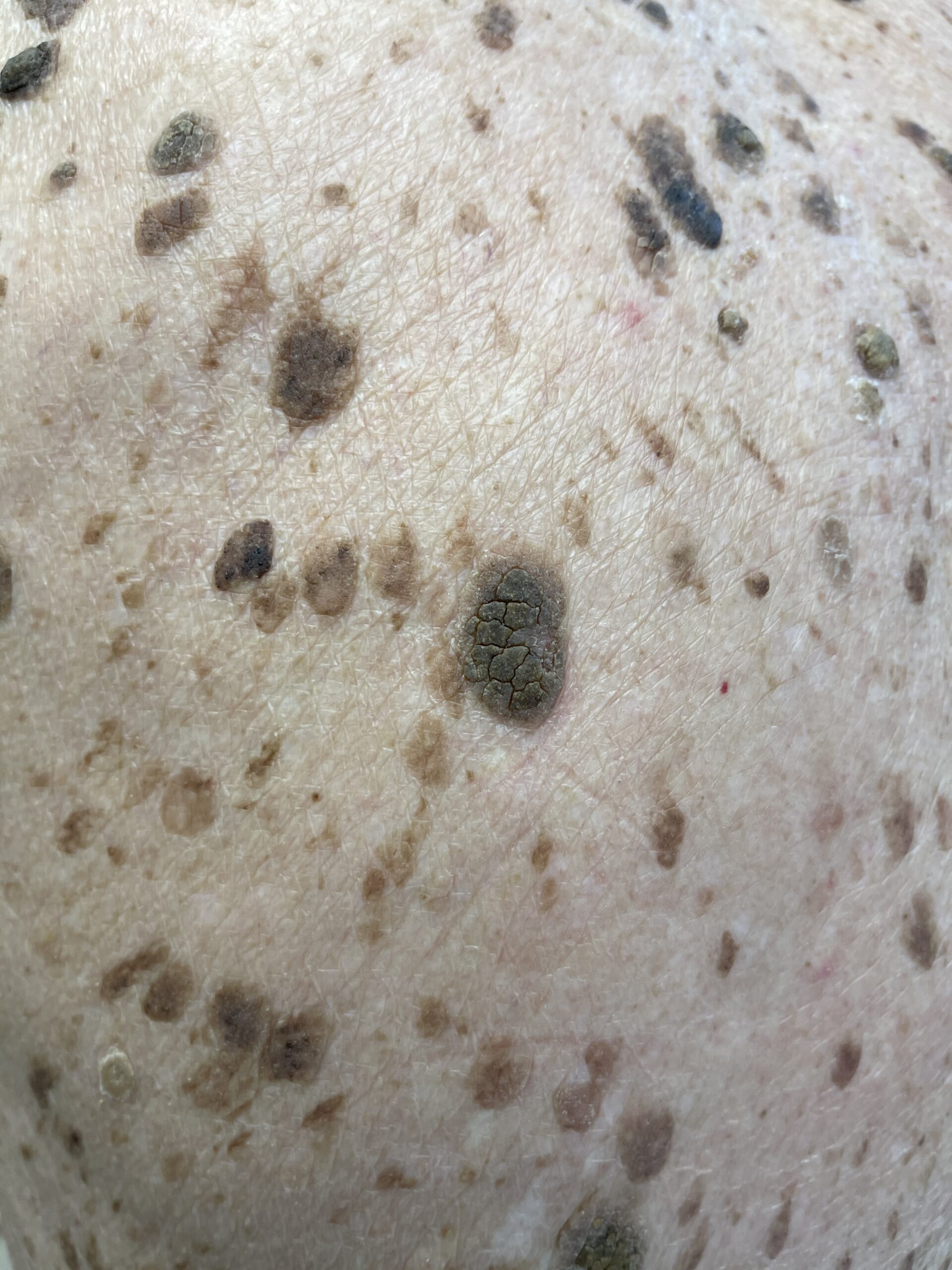
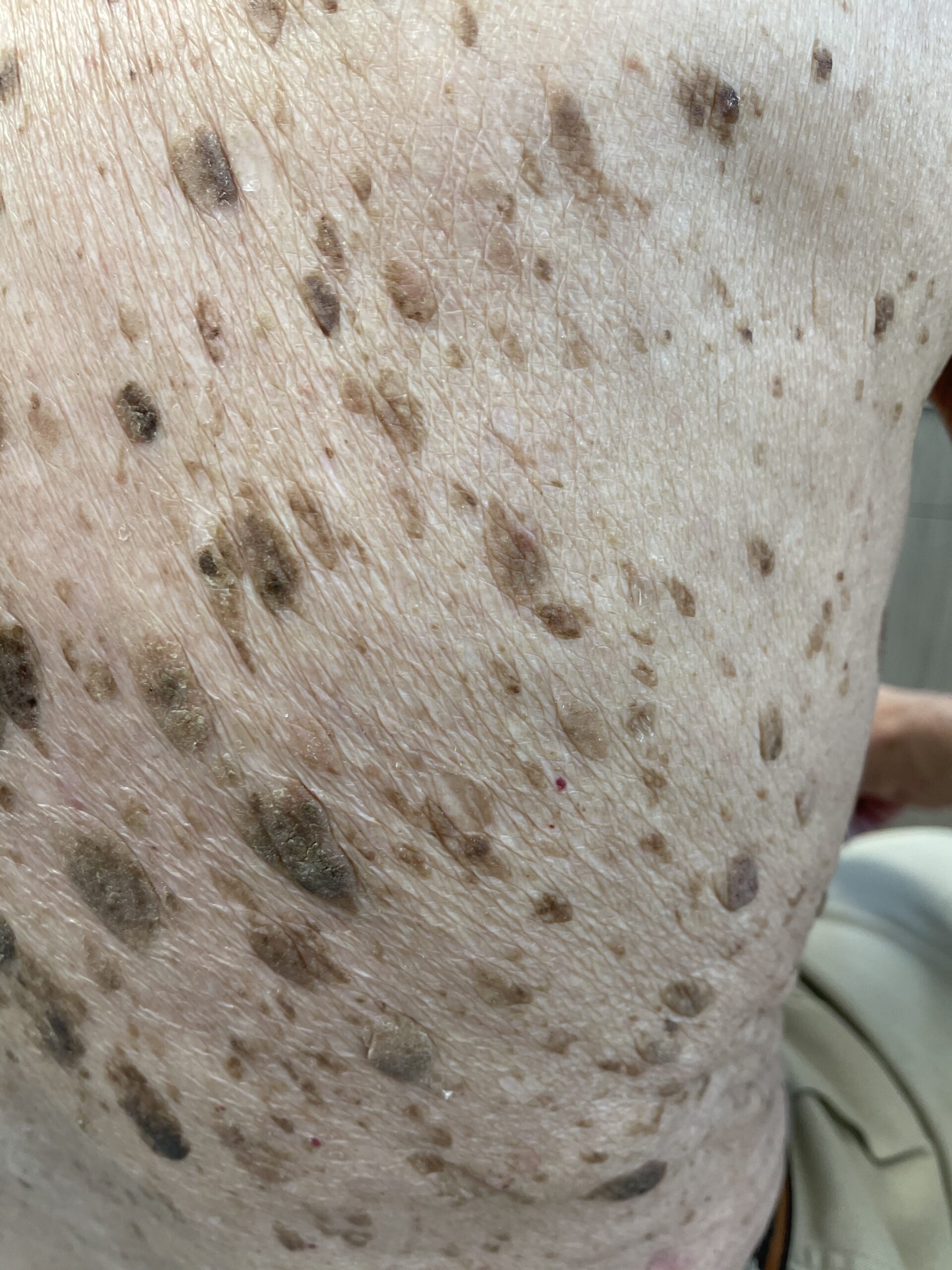
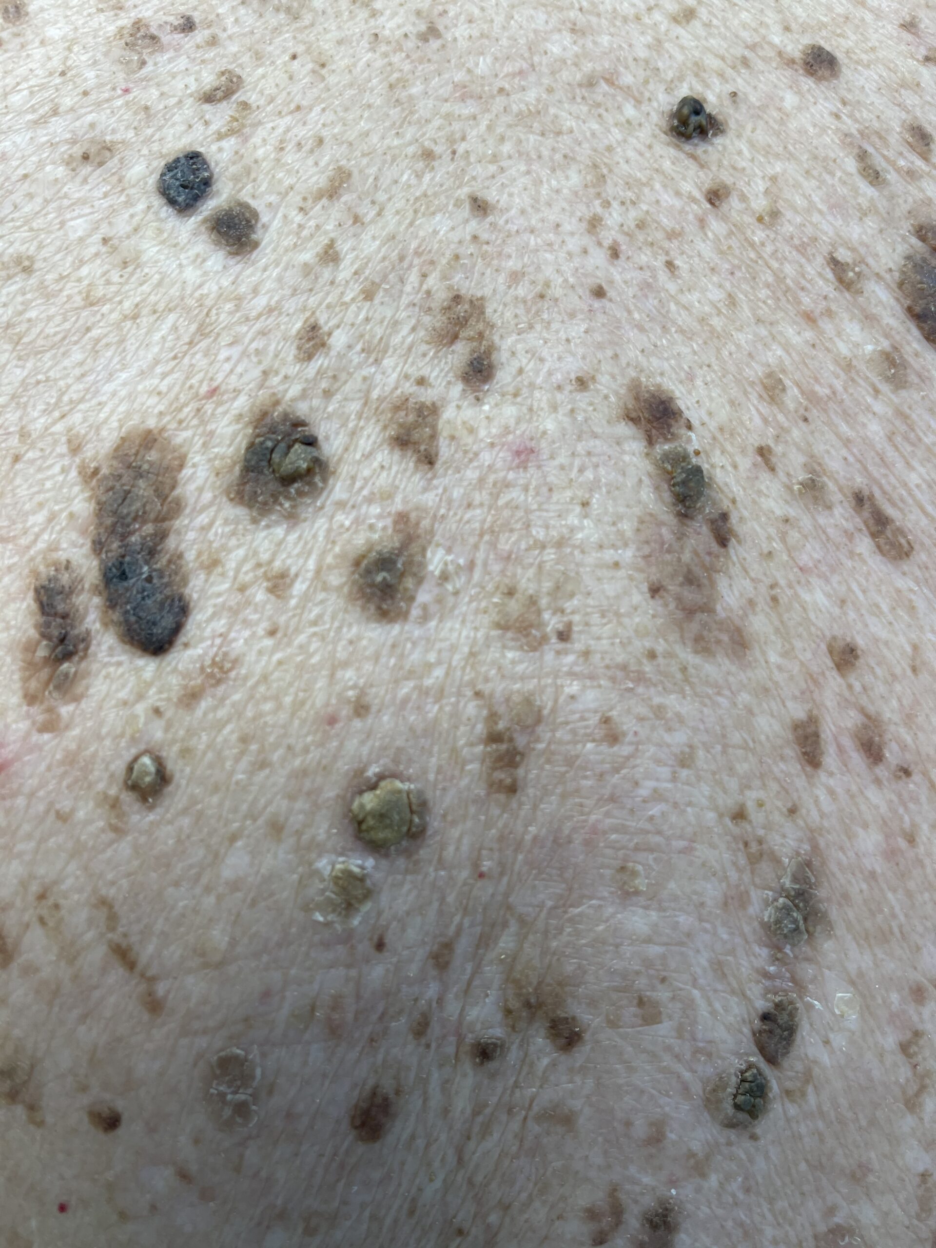
View the clinical video
When should a spot be assessed?
See a clinician experienced in assessing skin lesions if a spot is new, changing, bleeding, or uncertain. Some skin cancers can resemble seborrheic keratoses. A sudden appearance of many new growths should also be assessed.
Diagnosis
Clinical examination comes first. Your clinician looks at the growth and may use dermoscopy to examine it in more detail.
Dermoscopy
Dermoscopy uses illuminated magnification to reveal features that may support a diagnosis of SebK, such as small white or yellow cyst-like structures, dark plugs, and grooves or ridges. Some lesions do not show a typical pattern, and these features cannot always distinguish SebK from skin cancer. If uncertainty remains, a biopsy or removal for laboratory examination may be recommended.
What can dermoscopy show?
These are patterns a trained clinician may look for. They are not a checklist for ruling out skin cancer yourself.
- Cerebriform pattern — a brain-like surface of fissures and ridges
- Fingerprint structures — curved, parallel lines resembling a fingerprint
- Comedo-like openings — dark, blackhead-like plugs
- Milia-like cysts — small round white or yellow dots
- Hairpin blood vessels — looped vessels, often with a pale halo
- Moth-eaten border — a sharply scalloped, bitten-looking edge
Biopsy when needed
A biopsy is not needed for every typical SebK. If examination and dermoscopy do not establish a confident diagnosis, tissue may be examined by a pathologist before cosmetic treatment is considered.
Do they need treatment?
Once a clinician is confident that a growth is a seborrheic keratosis, it usually needs no treatment. An uncertain, changing, or bleeding lesion should be assessed before it is treated or removed.
Removal may be considered if a confirmed growth catches on clothing or jewellery, becomes irritated, or causes concern about its appearance.
Removal options
Freezing
Liquid nitrogen may be suitable for selected lesions, particularly thinner growths. Blistering and pigment changes can occur.
Curettage
The growth is scraped away, sometimes with an additional technique to control bleeding or treat the base.
Shave removal
Selected raised lesions may be removed with a blade. This can also provide tissue for laboratory examination when appropriate.
After removal
A treated area may form a scab or blister and usually heals over a few weeks. Follow the wound-care instructions provided by your clinician.
Changes in skin colour, either lighter or darker, can occur, particularly after freezing and in darker skin tones. Scarring is possible, and colour changes can sometimes persist. A growth may remain or recur after treatment, while new seborrheic keratoses can develop elsewhere with age. Complete clearance or a particular cosmetic result cannot be guaranteed.
Frequently asked questions
Is a seborrheic keratosis cancer?
No. It is a benign growth and does not turn into skin cancer. However, because melanoma can occasionally look similar, have any new or changing spot checked.
Are they contagious?
No. You cannot catch them or spread them to others, including family members.
Can I remove one at home?
No. Home removal risks infection, scarring, and — most importantly — missing a skin cancer. Any growth should be removed by a trained clinician.
Why do I keep getting new ones?
Gradual development of new growths is common with age and can run in families. A sudden appearance of many new growths should be assessed.
Unsure about a skin growth?
Contact Kelowna Private Skin Cancer Clinic to arrange assessment of a new, changing, or uncertain lesion. If it is confirmed as a SebK, you can discuss whether leaving it alone or removing it best suits your needs.